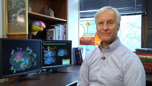
Hey everyone! So I have some really cool news! On Halloween, I am going to have my brain scanned with an unbelievable imaging technology called High Definition Fiber Tracking. This will be done at the University of Pittsburgh in Dr. Walter Schneider’s laboratory!
Many kinds of brain injuries (including concussions) can sever the axons, or nerve fibers, in the brain and body. These fibers act like phone lines to different areas, and if these cables are damaged, the communication between them is interrupted, which totally affects function. Comparing how we treat the body vs. how we treat the brain, if I walk into an emergency room with a broken arm and they treat my leg, it’s called malpractice. But it is common in the treatment of brain injury that there is a broken language cable, for example, but the treatment is for a broken visual cable.
We’ve all heard the saying: “If you’ve seen one brain injury, you’ve seen one brain injury.” We are very aware that each brain injury is different. The thing is that, until now, there was no diagnostic technique that could show us which axons are broken. Severed axons are often invisible on CT, MRI, or even functional MRI (fMRI), which measures brain activity by tracking blood flow. How are we to rehabilitate the approximately 1.7 million different people who suffer different TBIs each year in the US when we cannot tell what exactly is damaged? How can we rehabilitate different brain injuries when we don’t know what centers of the brain are not communicating with each other?
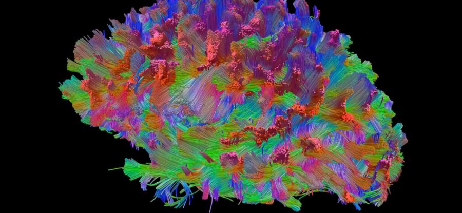
Enter High Definition Fiber Tracking. This imaging technique is apparently able to show exactly where these axon pathways are connecting! It can allegedly show the pathways along which these cables are traveling and where these connections are severed… and to do so non-invasively using MRI technology. That’s incredible! Are we now able to see exactly where the axons, or cables for communication, are severed? With this information, we should be able to target therapies to rehabilitate our brains functionality more than ever before! If we were to make a “Google Maps” of the brain, this kind of imaging is what we would need.
Did I mention that I’m being scanned on Halloween!?! 😮
As you can imagine, I’m very excited to explore this technology and to see if my brain is really being imaged in such vivid detail. You can learn more about it from this article in Discover Magazine, and if you have a brain injury and are interested in exploring it yourself, you may be eligible to have your brain scanned for free by visiting https://www.lrdc.pitt.edu/hdft/content/information.asp.
I’m so excited and will try to document as best I can while I’m in Pittsburgh. Wish me luck, and if you might be in the area and able to help out by holding a cell phone and filming, shoot me an email.
Cheers!
Cavin
*Blog Photo Caption: High definition fiber-tracking map of a million brain fibers. Credit: Walt Schneider Laboratory


9 Comments
Leave your reply.