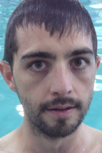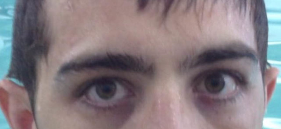
Notice how my right iris and pupil sits higher than my left. You can see the white below my iris on the right eye (left side of picture), but not on the left (right side of picture).
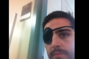
I have Diplopia or double vision. The image that I see with my right eye is below and to the right (outward) of the image that my left eye sees. Individually, each eye sees clearly, but the two images don’t line up when I use both. This is why I wore an eye patch for months, switching it from right to left eye every 20 minutes so I would not weaken either eye. I had 20×20 vision from the results of my eye exam a month before my accident and didn’t want to change that.
I had gone to see a doctor at SUNY optical while I was in NY in September, 2011 and she had prescribed a prism to shift the image that I saw with my right eye up seven diopters (visual units). I was using that lens for several months. When I first went to the Austin Center for Vision Development, I thought that my eyes were fine, but my brain wasn’t interpreting the images correctly.
It took me months to understand exactly what was going on with my double vision. I learned that it was and is yet another example of how my brain and body don’t communicate well. I was partially right that my eyes were fine, but my thought that my brain didn’t interpret it the alignment isn’t accurate. Diplopia is also often referred to as fourth nerve palsy. Normally, if you close your eyes for a while, and the open them and look at… say a penny, what happens is that your eyes see two separate images or two pennies (one from each eye) and immediately (a split second), your brain tells muscles in your eye to orient the eyes in a certain way so that the objects converge and you see the one penny that is actually there.
The fourth nerve controls the superior oblique muscle for eye movement, and palsy is a medical term meaning partial paralysis. The function of the superior oblique is primarily to move the eye downward and outward. Essentially the automatic eye movement to align my vision is partially paralyzed. You can see this in this photo. My right pupil is higher than my left… They are not at the same height.
Although the left side of my body is what is affected by my ataxia, my right eye was where the problem lay with my eye movement. The right side of the brain controls the left side of the body, yet it hooks up to the same side eye (right). It is a little strange to me, but evolution has a few examples of strange phenomena like this which actually support the theory of evolution. The recurrent laryngeal nerve (nerve that controls the voice box), for example, takes an extreme detour (about 15 feet in the case of giraffes) before it supplies motor function and sensation to the larynx (voice box). It is cited as evidence of evolution as opposed to intelligent design.
Sorry… I went on a tangent.
When I went to be assessed for my vision I was put through many tests. Dr. Denise Smith assessed my right eye as seeing nine diopters below my left eye. I had already deteriorated two diopters and in fact, the higher the prism, the more my brain liked it. The way this was tested was by switching the prism one unit up each time and asking me which one looked better. I said that the eight looked better that the seven, the nine looked better than the eight… and so on. Dr. Smith said said that I was “eating up the prism”, meaning that my brain was more than willing to get lazy and let the prism adjust my vision so that I no longer needed to use the paralyzed eye muscles. She said that if we don’t do something right now, that muscle will get lazier and lazier and not try to regenerate.
I was beginning to see double in my current prism strength and Dr. Smith had me stay in the seven prisms and begin therapy. On our way out the door, Dr. Smith said to my mom “I know this is expensive, thank you so much for caring enough to do this for him. I don’t know of he’ll ever not wear glasses, but I think we can make some real progress here.” I wasn’t happy with my vision in my current glasses, so I wasn’t happy with her plan to leave me in my current glasses because I saw double which made things frustrating, annoying, and dizzying. She instructed me to do a few exercises until I began therapy. I did just that, and miraculously saw my vision improve. The two images soon converged with a 7 diopter prism. This was still 7 units from where I need to be to see single, but things were finally moving closer together rather than further apart.
I am updating this post in light of Hillary Clinton’s testimony before the Senate Foreign Relations Committee on Wednesday January 23, 2013. Evidently she had fainted due to dehydration caused by a stomach virus. She lost consciousness, fell, and hit her head. She was diagnosed with a concussion on Dec 13, 2011. A common cause of fourth nerve, or superior oblique palsy is head trauma, including relatively minor trauma. A concussion or even whiplash may be sufficient to cause this problem.
If you look closely at this photo of Hillary, you will notice that there are small white lines running vertically on the left lens (right side of picture) of her glasses. These lines are from a stick on “Fresnel” prism. This is the same kind of prism that I had used before I had my prescription ground into my glasses.
The fact that she is using these Fresnel prism is evident that it will, hopefully, only be a temporary misalignment Perhaps she will do vision therapy in her copious amounts of spare time. If you know anything about Hillary Clinton’s position as Secretary of State at this time, you will know that I am being sarcastic.
SPOILER ALERT:
After a year of therapy, I currently see single in a four diopter prism! It’s working!
This is a section of my Keynote at the Neuro-Optometric Rehabilitation Associaion:


11 Comments
Leave your reply.